Transmission X-ray Microscopy
X-ray microscopes produces contrast using the difference in absorption of soft X-ray in the water window region.
Wavelength region: 2.3 – 4.4 nm, corresponding to photon energy region of 0.28 – 0.53 keV is where the carbon atom (main element composing the living cell) and the oxygen atom (main element for water) deliver good imaging contrast.
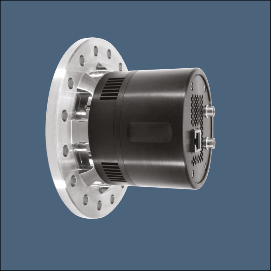
A typical set up consists of polychromatic source used with condenser optic that relays the radiation onto the sample and a Fresnel zone plate is used in order to magnify the image onto the camera. The latter is a very high resolution cooled high sensitivity sCMOS camera coupled to a state of art scintillator using high NA lens with 1.4 micron effective pixel size.
An alternative solution using direct exposure of the sCMOS to soft X-ray is also available with 11 microns pixel size.
An other technique, known as lens less coherent diffraction imaging is emerging as a potential technique for enhancing resolution down to 1.5 the radiation wavelength. It also enables to eliminate a low transmission FZP.
Combined with XANES (by taking an image above and below the absorption edge of an element), the camera can unveil information about the chemical state of components in the sample.
We Recommend...
X-Ray sCMOS 4MP Detector

Contact Us
22 Theaklen Drive,
Saint Leonards-on-sea,
TN38 9AZ,
United Kingdom


