TEM - Transmission Electron Microscopy
The TEM is used heavily in both material science / metallurgy and the biological sciences.
In both cases, the specimens must be very thin and able to withstand the high vacuum present inside the instrument.
Electrons are transmitted through an ultra-thin specimen, interacting with the specimen as it passes through it. An image is formed from the electrons transmitted through the specimen, magnified and focused by an electron lens and appearing as an image on a fluorescent screen coupled to a high-resolution CCD camera.
Spatial resolution and dynamic range are important because of the high intensity distribution across the image, especially during diffraction experiments. The ability to adapt speed of acquisition and dynamic range is required when acquiring fast structural data sets with cryogenically cooled samples.
Fibre optic coupling combined with proprietary scintillator deposition allows the optimum detective quantum efficiency and optimum resolution to be achieved from 70 up to >200 kV operation.
We Recommend...
X-Ray sCMOS 4MP Detector X-Ray sCMOS 16MP Detector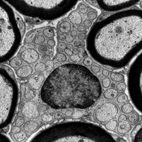
- Quality Policy
- Privacy Policy
- Terms of Use
- Export Compliance Statement
- Environmental Policy
- Conflict Minerals Policy
- Modern Slavery and Human Trafficking Statement
Certifications
We are ISO 9001 and 14001 certified by BSI.


Contact Us
22 Theaklen Drive,
Saint Leonards-on-sea,
TN38 9AZ,
United Kingdom