Diffraction Enhanced Imaging
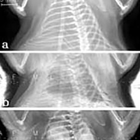
Diffraction Enhanced Imaging is used to unveil edge contrast in biological samples thanks to the detection of very subtle X-ray scattering changes.
An analyzer crystal is positioned between the sample and the image detector. The narrow angular acceptance of the analyzer allows to characterize the refracted and scattered X-rays going through the sample.
Scattering angles larger than the angular acceptance of the analyzer, typically few micro radians, will not be transmitted by the analyzer, this provides extinction contrast.
Equally, only the density variations across the sample that leads to micro radian refracted X-rays will go through the analyzer, this provides refraction contrast. Those subtle differences in scattering and refracting angles produced by the samples will be converted into intensity differences, which are then detected by a high resolution X-ray detector.
Sufficiently bright source must be used in order to collect enough flux after a low acceptance solid angle analyser, equally, a very high resolution detector must be used in order to sense very subtle edge contrast changes over a range of high spatial frequencies.
We Recommend...
X-Ray sCMOS 16MP Detector X-Ray sCMOS 64 MP Detector

Contact Us
22 Theaklen Drive,
Saint Leonards-on-sea,
TN38 9AZ,
United Kingdom


