Small Molecule and Protein Crystallography
This technique helps engineering of future drugs and chemical formulas produced by the pharmaceutical and chemical industry.
Small molecules (unit cell containing 100 atoms or less) and macromolecular (unit cell containing 10000 atoms or more) are crystallized and exposed to high brilliance X-ray beam on a synchrotron or X-ray lab source.
The experimental set up consists of gradually rotating the samples over 0.1 to 0.25 degrees in order to record Bragg reflections from each orientation of the crystal.
A good dynamic range is required, typically > 15,000:1 with potentially the possibility to read and expose at the same time in order to be able to rotate the sample at continuous speed over fine angular ranges: this is the fine Phi slicing technique.
Depending on beam delivery conditions as well as crystallization quality, the data collected can reveal conformal properties of a materialin addition to its electron density that will shed some light onto binding mechanisms of enzymes or proteins.
Large area detectors up 165mm diagonal detectors are more commonly used with laboratory sources.
We Recommend...
X-Ray sCMOS 16MP Detector X-Ray sCMOS 64 MP Detector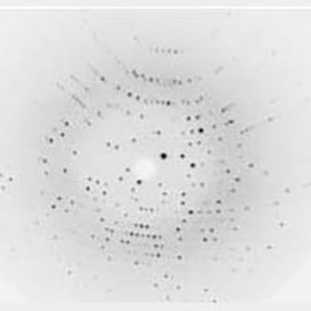
- Quality Policy
- Privacy Policy
- Terms of Use
- Export Compliance Statement
- Environmental Policy
- Conflict Minerals Policy
- Modern Slavery and Human Trafficking Statement
Certifications
We are ISO 9001 and 14001 certified by BSI.


Contact Us
22 Theaklen Drive,
Saint Leonards-on-sea,
TN38 9AZ,
United Kingdom