X-ray Micro and Nano tomography
X-ray Tomography allows a 3D reconstruction from a series of radiographs for different angular positions of the sample down to sub micron resolution.
Typically a full tomographic data set will require in the order of few hundreds to a few 1000s radiographs using a 3D reconstruction Feldkamp algorithm. Optical Cone beam / fan beam reconstruction are used, with the sample rotating in a fixed plan / helicoidally around an axis perpendicular to the beam.
The total acquisition time is in the range of few seconds per frame, it depends very much on the source brilliance / geometry. 100% duty cycle detectors with simultaneous read out / exposure allows to save up to 50% of the scanning time.
Resolution down to a few hundred nanometers can be achieved by using a small focal spot source and reasonable geometric magnification. The recorded data is often several Gigabytes and can be processed using the massively parallel calculation capacity of GPUs.
Micro tomography can be combined with phase contrast imaging, either in a qualitative way (“edge enhancement”) or, more quantitatively, including phase retrieval (“holotomography”). Very high resolution cameras allows the build of scanners with sub micrometer spatial resolution whilst keeping compact dimensions and good sensitivity.
We Recommend...
X-Ray sCMOS 4MP Detector X-Ray sCMOS 16MP Detector X-Ray sCMOS 64 MP Detector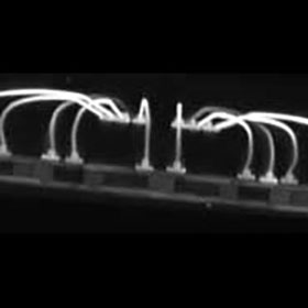
- Quality Policy
- Privacy Policy
- Terms of Use
- Export Compliance Statement
- Environmental Policy
- Conflict Minerals Policy
- Modern Slavery and Human Trafficking Statement
Certifications
We are ISO 9001 and 14001 certified by BSI.


Contact Us
22 Theaklen Drive,
Saint Leonards-on-sea,
TN38 9AZ,
United Kingdom