Small Animal Imaging
Luminescence emissions from reporter genes provide a quantitative model for studying the development of human diseases. As the amount of light collected using this method is very little, an ultra-sensitive camera is required to record luminescence emissions as they propagate through soft tissues.
PSEL cameras offer very low read out noise, down to a couple of electrons, with negligible dark current that allows the capture of very low photonic emissions, typically down to few hundred photons per second per steradian and cm2 and over minutes of exposure.
These measurements are usually combined with fluorescence and X-ray CT scans in order to provide an accurate 3D model of tumour / location propagation. To further improve image contrast at increased tissue depth, much attention has been focused on the development of NIR-I-to-NIR-II fluorescence imaging, which can remarkably reduce the interference from photon absorption, scattering and tissue autofluorescence with excitation in the 700–950 nm NIR-I window and emission in the 1000–1700 nm NIR-II window.
Reference Articles
Real-time in vivo visualization of tumor therapy study incorporating SWIR camera from PSEL
We Recommend...
Cooled VGA SWIR InGaAs Camera X-Ray FDS Detector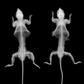
- Quality Policy
- Privacy Policy
- Terms of Use
- Export Compliance Statement
- Environmental Policy
- Conflict Minerals Policy
- Modern Slavery and Human Trafficking Statement
Certifications
We are ISO 9001 and 14001 certified by BSI.


Contact Us
22 Theaklen Drive,
Saint Leonards-on-sea,
TN38 9AZ,
United Kingdom